longitudinal cross section of kidney
Anatomy of the Kidneys. 11 Pictures about Anatomy of the Kidneys : Cross Section of Right Kidney - Stock Image - C024/9532 - Science Photo, Renal Pathology and also Kidney Tubule Under Microscope - kidneyoi.
Anatomy Of The Kidneys
 www.brainkart.com
anatomy kidneys structure nephron kidney location describe parts
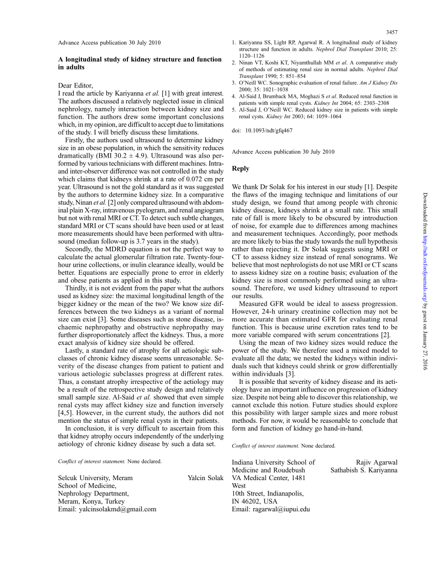
(PDF) A Longitudinal Study Of Kidney Structure And Function In Adults
 www.researchgate.net
longitudinal
Urinary System Disorders | Basicmedical Key
 basicmedicalkey.com
structure renal system urinary function kidney disorders section mosby coronal st figure adapted anatomy internal lobe structural urologic systems nursing
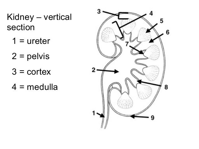
Kidney – Structure And Function
 www.slideshare.net
renal ureter cortex medulla glomerulus
Chapter 14, Page 2 - HistologyOLM
 stevegallik.org
esophagus histology histologyolm chapter source gi acm uiuc edu project stevegallik
Kidney Tubule Under Microscope - Kidneyoi
 kidneyoi.blogspot.com
cellula epiteliale epithelial kidney eenvoudige intestinale rene cellule epiteliali tubule niere umano coggle organisme diagram intestinal pictogramreeks wetenschaps pictogramembleem deoxyribonucleic
Home.php Lab
 histology.med.yale.edu
kidney histology urinary ducts renal pelvis bladder ureter connects
| (A) Cross-section Of The Cochlear Duct, Illustrating The
 www.researchgate.net
cochlear illustrating
Renal Pathology
 library.med.utah.edu
kidney normal renal kidneys section cross cortex pathology anatomy adult medulla medical collecting webpath utah med edu views darker lighter
Cross Section Of Right Kidney - Stock Image - C024/9532 - Science Photo
 www.sciencephoto.com
Human Simple Cuboidal Epithelium Single Kidney Tubule Also Shows Lumen
 www.gettyimages.com
epithelium cuboidal simple kidney tubule lumen human section tissue single longitudinal mallory stain surrounding shows connective res collecting duct x250
Kidney normal renal kidneys section cross cortex pathology anatomy adult medulla medical collecting webpath utah med edu views darker lighter. Kidney – structure and function. Esophagus histology histologyolm chapter source gi acm uiuc edu project stevegallik